

We will attempt to outline the differences between two very different types of images compared to the photographs we are used to seeing daily in newspapers, magazines, or with our own eyes:
Multispectral Images and Hyperspectral Images
First, let’s analyze the words “image” and “spectrum” separately:
- Image: Reproduction of an object’s figure by the combination of light rays emanating from it.
- Spectrum: Distribution of the intensity of radiation as a function of a characteristic magnitude, such as wavelength, energy, frequency, or mass.
Therefore, a spectral image is one that reproduces the figure of an object based on the wavelength the object in question is reflecting (or emitting); or, in other words, it is a set of images of the same object, each represented with different wavelengths.
The differences between a multispectral image and a hyperspectral image are numerous, but the main one is the number of spectral bands. The following image perfectly illustrates this difference:
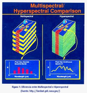
We can say that multispectral images are composed of relatively few bands (usually between 3 and 20) and these bands are not necessarily contiguous, whereas hyperspectral images are typically composed of a larger number of bands, and these are always contiguous.
With a multispectral image, we can obtain intensity values at the discrete wavelengths where the system captures radiation, while with a hyperspectral image, we obtain the continuous spectrum or spectral signature of the analyzed object.
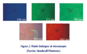
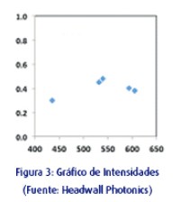
The following images were captured with a multispectral camera using spectral filters at 436, 532, 540, 594, and 605nm ± 20nm (false color representation), Figure 2. If we normalize the intensity and plot the values of the same pixel across the 5 images in a graph, the result would be that shown in Figure 3.
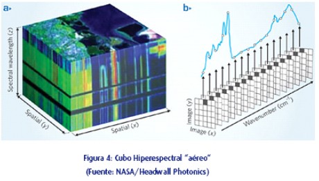
Let’s look at a hyperspectral image (false color representation) with the corresponding continuous spectrum at one pixel of the image:
The way multispectral and hyperspectral images are captured also differs greatly.
While multispectral cameras are frame-type and capture images with 2 spatial dimensions (X, Y), pushbroom hyperspectral imaging systems generate “images” with 1 spatial dimension and one spec
tral dimension (X, Z), which corresponds to the left side face of the cube shown in Figure 4.
The process to obtain a hyperspectral cube involves scanning the scene to generate the second spatial dimension, and this scan can be performed in two ways: by moving the object or by moving the hyperspectral camera over the scene or the object of study.
Regarding the use of one type of image or the other, we can say that hyperspectral images offer much more quantitative information and are used as tools for spectral differentiation and classification, whereas multispectral images contain less information but are very useful when we know the wavelengths
that differentiate one material from another.
